SHARE WITH FRIENDS:
Pelvic joint dysplasia is a congenital disorder of the formation of the pelvic joint, the complication of which is the protrusion of the head of the hip. In this case, the joint is not fully developed or there are not enough structural elements made of connective tissue. At an early age, skin folds are characterized by asymmetry, relative shortness, and limitation of hip mobility.
Later, there are cases of pain, lameness, and fatigue. The pathology is diagnosed by UTT examination and radiography. Therapeutic procedures are performed using special fixatives and special exercises.
Pelvic dysplasia (Greek dys - disorder, placeo - formation) Is a congenital pathology, complicated by the protrusion or partial protrusion of the head of the thigh. The degree of non-formation of the joint is simply a cosmetic inconvenience or a variety of movement defects. To avoid complications of pelvic dysplasia, the pathology should be treated early, ie from the first month of life.
This pathology is the most common congenital disease, accounting for 2-3% of all births. Among ethnic groups, it is less common among African Americans and more common among American Indians than among other races. Girls are more likely than boys to suffer from pelvic dysplasia (80% of all pathologies).
Causes of pelvic dysplasia
Several factors are involved in the origin of dysplasia. Hereditary factors, such as the presence of congenital pelvic pathologies in the parents, increase the risk of the disease by up to 10 times. In addition, pelvic fetal delivery is one of the leading causes of pathology. Pregnancy timely toxicosis, taking various drugs, fetal size, lack of fetal fluid and other pregnancy pathologies are also noted to cause dysplasia.
Researchers have found that babies born with pelvic dysplasia are five to six times more likely to be born in environmentally unsatisfactory areas. Pelvic floor dysplasia also depends on the customs of the nation, that is, children are diagnosed differently in different regions. For example, this pathology is less common in areas where the cradle is open between the legs or where children are carried with their legs open in the back.
Pathogenesis
The pelvic joint consists of the femoral head and the pelvic floor. The upper part of the pouring bowl consists of the pouring lip, and this part expands the area as the head of the thigh enters the cup. The pelvic joints of newborns are different from those of adults: the pelvic floor is flat, almost vertical, and the elbows are more elastic. The head of the thigh is attached to the pelvic girdle by a round neck, a joint capsule, and the labia minora.
There are three types of pelvic dysplasia: acetobular (defects in the development of the pelvis), dysplasia of the upper femur, and rotational dysplasia, which is a change in the geometry of the bones on a horizontal surface.
Disruption of any of the elements that make up the joint results in the inability to ensure that the head of the joint is in the cup. As a result, the head of the number protrudes outwards and upwards. The bowl also changes its structure and loses the ability to attach the head of the number. If the head of the thigh is completely out of the bowl, the head of the thigh is at the top. If not treated in time, the pelvic girdle fills with connective tissue and adipose tissue, making it difficult to replace the head of the thigh.
Symptoms of dysplasia
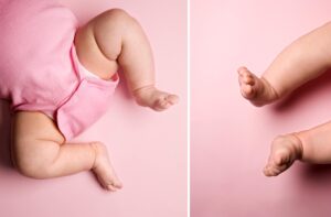
Pelvic dysplasia is the first step in diagnosing pelvic dysplasia based on the relative shortness of the legs, asymmetry of the skin folds, limited range of motion, and Marks-Orthola. Asymmetry of skin folds in the groin, back of the knee and buttocks areas is mainly well known in children aged 2-3 months. The vision focuses on the differences between the folds, the shape and the depth of the folds.
The above tests are not sufficient to diagnose dysplasia. In cases of bilateral dysplasia, the folds may also be symmetrical. Occasionally, asymmetry of skin folds can be observed in healthy children. The relative shortness provides good information for determining the birth rate. When the child is lying on his back, his legs are bent at the pelvis and knee joints, and attention is paid to the position of the knees relative to each other, if one side is above the other - this indicates a congenital hip dislocation.
The Marx-Orthodox sign gives the most accurate information in determining the origin of a congenital number. The child is placed on his back, the doctor bends the child's leg and holds it with his hands so that the fingers II-V are located on the outer surface of the thigh. The doctor then bends the thigh out at the same time with the same force. In the case of dysplasia, on the pathological side, a squeaking sound is heard due to the return of the head of the thigh to the pelvic floor. This symptom is positive in 1% of newborns in the first week of life and then disappears on its own.
There is another way to diagnose joint pathology - limited mobility. In healthy children, the legs open outwards to 80-90. When movement is limited, this angle is 50-60 percent.
Complications of pelvic joint dysplasia
Arthral dysplasia may not cause any complaints in young children. However, dysplastic when a person reaches the age of 25-55 years coxarthrosis are more likely to develop. The first symptoms may be limited mobility or hormonal changes in women.
The peculiarity of dysplastic coxarthrosis is that the disease begins in a very short time and progresses rapidly. The disease is accompanied by discomfort, pain and limited mobility in the joints. In the late stages, the legs become protruding, bent. Movement in the joint is severely limited. The head of the thigh rises slowly and forms a false joint in the femur, but this is very rare today.
Diagnostika
Pelvic joint dysplasia is diagnosed in the maternity ward. If pathology is detected, a pediatric orthopedist should be consulted within 3 weeks. In addition, children are screened at the age of 1, 3, 6 and 12.
Children at risk, for example, in the mother during pregnancy anemia, toxicosis, fetal size, pelvic placement, and children born to parents suffering from dysplasia should be considered. If dysplasia is suspected, the necessary tests should be performed.
Radiographic and ultrasonographic examinations are performed to confirm the diagnosis. In young children, the joint elements are made up of ridges and are not visible on the radiograph. Therefore, radiography is performed in children older than 3 months. Ultrasonography is very convenient and harmless for young children.
Diagnosis is made with instrumental examination results and a general examination
Treatment of pelvic joint dysplasia
Treatment should be carried out at an early stage. It uses a variety of fasteners that hold the child's legs open and bent: devices, tires, straps, barbells and special pads.
It is better for young children to use devices with elastic construction. For example, the Pavlikov stremen is a device for attaching to the chest, in which the baby's legs are hung out on the mother's chest with the legs outstretched and bent at the knees. Another advantage of this device is that the movement of the child's legs is not limited.
One of the most effective ways to treat pelvic dysplasia is to perform special exercises that strengthen the muscles. In addition, buttock muscle massage is also prescribed.
In severe cases, a plaster cast is applied and the joint is immobilized while keeping the legs in the desired position. This type of treatment is given to children between the ages of 2 and 6. Children under the age of 8 are counted. If these measures do not work, the head of the thigh is lowered to the surface of the joint with surgery.
Consequences and prevention of pathology
As a result of early detection of pathology and initiation of treatment, the outcome of the disease is positive. The later the disease is treated, the higher the risk of complications. Prevention of pelvic dysplasia involves early detection of pathology in children.